 |
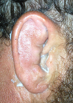
| Patient 1 |
This is a picture of some guy’s right ear. We'll call him Patient 1. For simplicity’s sake, I’ll italicize medical
terminology between parenthesis. The ear
obviously doesn’t look right (abnormal). It’s swollen (edematous), and when you squish it about between your fingers (palpate it), it surprisingly feels as if
there is some fluid deep to it, as it is quite mushy (fluctuant) and altogether entirely unsettling.
He tells me this occurred about a week and a half ago, after
hitting it hard against the water while wake-boarding on the river, being
pulled by a fast-moving boat. He went to
the emergency room a few days later since it started to swell even more and was
given some antibiotics. He went back to
the ER since it wasn’t getting better after a week, and was told to see an ENT
doc and thus the reason he came to my office.
He has an auricular hematoma. Hematoma is a collection of blood. It is somewhat of a misnomer, as -oma in medical jargon means “tumor”,
but this is not really a tumor. Hema refers to blood. So this technically would be a “blood tumor”
which again isn’t entirely the case, but more of a collection of blood in a
space that shouldn’t exist. In this
case, the blood occurred from ear trauma, causing bleeding to occur between the
cartilage and the perichondrium—a tight layer of tissue covering the cartilage. This causes a mass per se, and on the ear it
is quite visible. All of this lies deep
to the skin. The mass effect doesn’t go
away by itself. Later, the hematoma
“organizes, meaning the blood cells break down, and what remains is the serum,
or liquid that transports blood cells through the body. When this occurs, the mass becomes a seroma.
A Cauliflower Ear
is the deformity of the cartilage if this problem is left untreated. The perichondrium provides the blood supply
to the cartilage, and when this is disrupted by something as a hematoma/seroma,
parts of the cartilage will be undernourished.
It will tend to scar (fibrose)
and then contract, causing a deformity of the cartilaginous framework that
gives the ear its shape.
This phenomenon often occurs in wrestlers, where the ear is
grabbed during a wrestling bout. This is
the reason wrestlers should wear ear protection. I’ve also seen it in mixed martial arts
fighters who, like a lot of guys in general, leave it untreated and thus
creating the Cauliflower look. Patient
2 below is a guy
whose ear was grabbed during a jiu-jitsu match.
As it so happens, this particular patient wrestled in high
school years ago and had this same problem back then, only at that time the
hematoma was much smaller, such that a doctor or coach or his Uncle Ernie (he
didn’t recall details) drained it by poking it with a needle and draining off
the fluid (aspiration), doing this
daily over a course of several days. Eventually
it resolved. So, on this occasion he
took it upon himself to purchase a box of sterile needles and treat it himself. He admitted to draining a bunch of fluid but
the darn mass recurred by the next day, even though he did the self-needing
over several consecutive days.
With this type of problem, the tissue still secretes serous
fluid once it is drained, filling up the space again. Sometimes very small hematomas will heal and
scar down such that the space is eliminated, but larger ones will fill back up
with fluid unless external pressure is applied to flatten the space and allow
the opposing tissue interfaces to bind to one another and seal, thus
eliminating the space and preventing re-accumulation of fluid.
We typically perform an incision and drainage (I & D) and
afterwards secure bolsters—soft pads that apply constant pressure on each side
of the drained space—to prevent recurrence.
“Ooh! How’s this
done?” I see you raising your hand with
impatient curiosity. So for you
Do-It-Yourselfers, below is a short synopsis, for interest-sake only, and as
always a warning: don’t DIY, even if you’re a medical professional. Bad things invariable happen. In other words, don’t be stupid and see a
doctor.
1. The doc must numb
(locally anesthetize) the ear. This requires injecting something like
lidocaine behind the ear first to numb the nerves that innervate the ear from
behind, and then over the top of the planned incision. Usually epinephrine is added to the lidocaine
to minimize bleeding when the incision is made.
2. After cleaning and
prepping the ear with a sterile cleanser, an incision is made paralleling the natural
contours of a normal ear, usually in a slightly curved line (curvilinear fashion in surgeon’s
jargon). If this is done a week or more
after the initial injury, a ton of amberish looking (serous) fluid pours out. If
this is done a few days after, then old blood and blood clots are often encountered. Once all of the fluid or old blood 1s removed
(evacuated), the ear’s appearance
usually is much improved, and the more normal cartilaginous shape is then
appreciated.
3. The evacuated
space is probed for any residual blood clots and sometimes irrigated with a
sterile salt solution (normal saline).
4. Bolsters are then
sewn through the ear.
Yes, the sutures
are placed through the skin, the underlying cartilage, and then out through the
skin on the other side. Bizarre, I know,
but this works well. A bolster is placed
on each side in order to apply pressure from both sides, sort of like an ear
sandwich so to speak. The sutures are
tied down rather tightly. There are other methods of applying pressure, such as
plaster, firm bandaging, but this technique seems to work the best. Often multiple bolsters are needed as
demonstrated in this case. |
| Patient 1: bolsters after incision and drainage |
5. A week later, the
sutures are cut and the bolsters removed. This provides ample time for the
tissues of the ear to properly heal and begin scarring down. The patient is instructed not to manipulate
or tug on the ear for another 3-4 weeks to prevent recurrence: for Patient 1, no wake-boarding and for Patient 2, no ear-grabbing.
 |
| Patient 1: after bolster removal one week later |
 |
| Patient 2: after bolster removal |
So there you have it.
Hope the curiosity is satisfied for those who were curious in the first
place. Again, no DIYs please, since we
don’t want to see you hurting yourself, as if smashing your head against the
water wasn’t bad enough.
©Randall S. Fong, M.D.


Comments
Post a Comment